THERYA NOTES 2026, Vol. 7:115-119
Record of unilateral ocular anomaly in a yellow-shouldered bat Sturnira hondurensis (Chiroptera: Phyllostomidae)
from Nauzontla, Puebla, México
Registro de anomalía ocular unilateral en un murciélago
Sturnira hondurensis (Chiroptera: Phyllostomidae)
en Nauzontla, Puebla, México
Gihovani Ademir Samano-Barbosa1, Angelica Maribel Orozco-Robles1, Miguel Ángel Aguilera-García2 and Miguel Ángel León-Galván3*
1Doctorado en Ciencias Biológicas y de la Salud. Universidad Autónoma Metropolitana. Iztapalapa. Av. San Rafael Atlixco No. 186, Col. Vicentina, CP 09340. Ciudad de México, México. gihovanisamano@gmail.com (GASB), angieorzc3542@gmail.com (AMOR).
2Maestría en Biología. Universidad Autónoma Metropolitana. Iztapalapa. Av. San Rafael Atlixco No. 186, Col. Vicentina, CP 09340. Ciudad de México, México. miguelaguileragarcia7@gmail.com (MAAG).
3Departamento de Biología. Universidad Autónoma Metropolitana. Iztapalapa. Av. San Rafael Atlixco No. 186, Col. Vicentina, CP 09340. Ciudad de México, México. leon@xanum.uam.mx (MALG).
*Corresponding author
Ocular anomalies in wild bats are poorly documented, especially under natural conditions. This report presents a case of unilateral ocular anomaly in an adult male Sturnira hondurensis. The individual was captured incidentally on May 16, 2025, in a cave in Nauzontla, Puebla, México, using a mist net. The individual was measured, photographed, and subsequently released alive at the capture site. During the physical examination, a translucent, gelatinous protrusion was observed completely covering the left eye, while the right eye showed no visible alterations. The appearance suggests a possible corneal alteration. This uncommon finding highlights the importance of including basic ophthalmological assessments in field studies, especially in areas where exposure to environmental contaminants may affect visual health. Documenting such anomalies will contribute to future clinical and ecological research on bats.
Keywords: Cave, eye anomaly; Phyllostomidae; visual health; wildlife
Las anomalías oculares en murciélagos silvestres están poco documentadas, especialmente en condiciones naturales. Este reporte presenta un caso de anomalía ocular unilateral en un macho adulto de Sturnira hondurensis. El individuo fue capturado de manera incidental el 16 de mayo de 2025 en una cueva en Nauzontla, Puebla, México, mediante una red de niebla. El individuo fue medido, fotografiado y liberado vivo posteriormente en el sitio de captura. Durante el examen físico se observó una protuberancia translúcida y gelatinosa que cubría completamente el ojo izquierdo, mientras que el derecho no mostraba alteraciones visibles. La apariencia sugiere una posible alteración corneal. Este hallazgo poco común destaca la importancia de incluir evaluaciones oftalmológicas básicas en estudios de campo, especialmente en zonas donde la exposición a contaminantes ambientales podría afectar la salud visual. La documentación de estas anomalías contribuirá a futuras investigaciones clínicas y ecológicas en quirópteros.
Palabras clave: Anomalía ocular; cueva, fauna silvestre; Phyllostomidae; salud visual
© 2026 Asociación Mexicana de Mastozoología, www.mastozoologiamexicana.org
Bats (Order Chiroptera), with approximately 1,500 species (Simmons y Cirranello, 2025), represent one of the most diverse and ecologically significant groups of mammals. Currently, Chiroptera is divided into around 21 families, which vary widely in species richness, from families with only a few species to highly diverse families such as Vespertilionidae and Phyllostomidae (Simmons and Cirranello 2025), exhibit adaptations to a wide range of ecosystems and diets (Teeling et al. 2018). The family Phyllostomidae, endemic to the Americas, comprises 230 species in 61 genera (Simmons and Cirranello 2025). Many of these are frugivorous and play an active role in seed dispersal (Shilton et al. 1999; Solari et al. 2019). Due to their ecological importance and diversity, phyllostomid bats are among the most extensively studied groups, which has allowed a relatively detailed understanding of their morphology and physiology, including sensory systems such as vision.
Although the ocular anatomy of bats has been described in certain species (Prince 1956; Blackwood et al. 2010), studies on visual health particularly under wild conditions are scarce. The research of Liu et al. (2015), has advanced our understanding of the molecular and electrophysiological mechanisms of bat vision, highlighting its functional importance despite the reliance on echolocation. Ocular conditions are most frequently reported in captive or rehabilitating individuals (e.g., DiGeronimo et al. 2018; Turner et al. 2021), while records from free-ranging bats remain exceptional, including in Mexico, where only a few isolated reports have documented such conditions in wild populations (Sánchez-Hernández et al. 2018). This underreporting may be due to the difficulty of detection or reduced survival of affected individuals (Montiani-Ferreira et al. 2022).
Sturnira hondurensis (Chiroptera: Phyllostomidae) is a frugivorous bat belonging to the subfamily Stenodermatinae, widely distributed from southern Mexico through Central America to northern South America. It inhabits a variety of environments, including tropical evergreen and semi-deciduous forests, as well as disturbed habitats (Hernandez-Canchola et al. 2021). Despite being studied in terms of its ecology and distribution, information on pathological conditions in this species is scarce. To date, there are no reports specifically documenting ocular anomalies in S. hondurensis across its distribution. The aim of this report is to present a case of unilateral ocular anomaly in a S. hondurensis bat from Nauzontla, Puebla, Mexico.
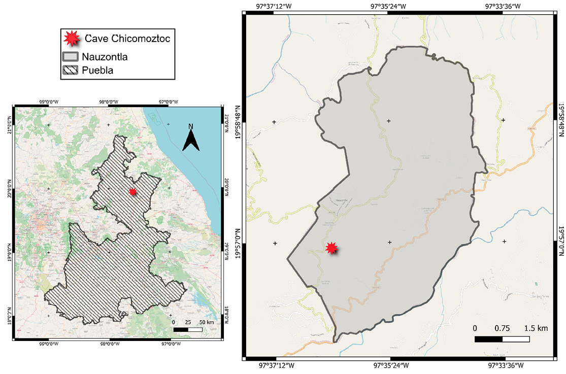
A nocturnal survey was conducted on May 16, 2025, in Chicomostoc Cave, located in the municipality of Nauzontla, Puebla, Mexico (Figure 1; 19° 56’ 55.4” N, 97° 36’ 18.9” W; 1,420 m), an area characterized by a humid semi-warm climate with rain all year round, and an average annual ambient temperature of 20 °C. Agriculture is an important economic activity in this region, with the main crops being potatoes, corn and beans, these crops are grown among grasslands and patches of cloud forest (SPF 2025). The present record occurred during field sampling conducted as part of a study aimed at evaluating the ecotoxicology of Myotis velifer. The study was conducted in accordance with established ethical guidelines (Sikes et al. 2016) and under permit SPARN/DGVS/13548/24 issued by the Dirección General de Vida Silvestre of SEMARNAT, Mexico, granted to Dr. Luis Manuel Guevara Chumacero.
A mist net was placed at the entrance of the cave to capture bats, and the species recorded were identified using specialized field guides (Sánchez-Hernandez et al. 2016; Alvarez-Castañeda et al. 2017). For each captured individual, sex and standard morphometric measurements (weight and forearm length) were recorded. Age was assessed based on the degree of ossification and closure of the metacarpophalangeal epiphysis of the fourth finger (Kunz and Anthony 1982). During the sampling, an adult male S. hondurensis was captured incidentally, as it was not part of the primary study objective. Photographs of the individual were taken in lateral, frontal, and ventral views using a Canon EOS Rebel T7 camera. The bat was subsequently released alive at the same capture site.
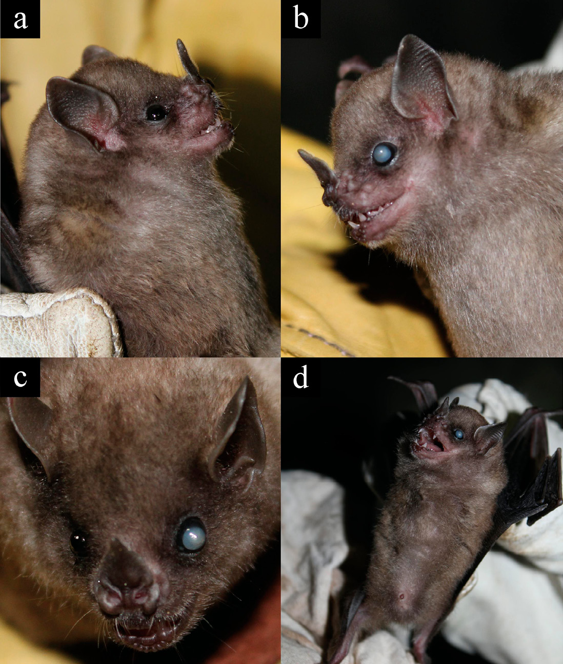
During the external inspection of an adult male S. hondurensis (weight: 22.3 g, forearm: 44.2 mm), a unilateral ocular anomaly was detected. The left eye exhibited a spherical, translucent protrusion with a smooth, gelatinous appearance, completely covering the eyeball (Figure 2). The right eye appeared normal. No other external abnormalities were observed, and the individual was active during handling.
Ocular anomalies may result from genetic predisposition, intraocular inflammation, senile degeneration, trauma, toxicity, environmental factors such as prolonged exposure to certain types of light, or systemic diseases, typically inflammatory or metabolic in nature (Turner et al. 2021). In bats, these conditions have been documented across different families and geographic regions worldwide. For example, cataracts and age-related degenerative changes have been frequently reported in captive species of the family Pteropodidae in Africa, Asia, and Oceania (Turner et al. 2021). In contrast, reports from wild Neotropical bats, particularly within the family Phyllostomidae, are scarce and mostly limited to isolated cases. In Mexico, Sánchez-Hernández et al. (2018) reported several ocular anomalies in free-ranging bats, including an adult female Artibeus lituratus with unilateral corneal opacity in the right eye, and an adult male of the same species with total corneal opacity and an enlarged eyeball in the right eye. Additionally, an adult male Sturnira parvidens presented a lesion in the left eye characterized by corneal perforation, edema, and purulent discharge, while the right eye appeared normal. In general, ocular conditions reported in bats worldwide include conjunctivitis, chemosis, and ocular discharge, primarily documented in captive or rehabilitated individuals (Montiani-Ferreira et al. 2022). Although no pathological or ophthalmological examination was performed to confirm the etiology, the macroscopic characteristics of the anomaly such as specifically the spherical, translucent protrusion and the loss of corneal transparency, suggest clinical signs compatible with corneal alterations or severe ocular distension, as described in other Neotropical bats such as A. lituratus and S. parvidens (Sánchez-Hernández et al. 2018; Montiani-Ferreira et al. 2022). While a definitive diagnosis is not possible without specialized clinical tools, the external morphology observed is consistent with the presentation of exophthalmos or corneal edema (Montiani-Ferreira et al. 2022). However, current evidence does not support a higher predisposition to ocular anomalies in any particular bat family or genus, including taxa closely related to S. hondurensis, as available reports remain scarce and largely anecdotal.
In this regard, cataracts have been observed in a significant number of captive individuals from various pteropodid species, most of them without an identified underlying cause. However, most cases involved middle-aged or older individuals (Turner et al. 2021), making age-related ocular changes a less likely explanation in this case, as the individual reported here was an adult. Additionally, Turner et al. (2021) considered toxicity an unlikely cause in captive pteropodids due to the controlled conditions in which the bats were maintained. In contrast, the study site corresponds to a natural cave environment surrounded by temperate forest vegetation and agricultural areas, where bats are exposed to natural environmental conditions. Although no direct evidence of contamination was identified during sampling, the potential influence of environmental factors, including toxic agents, cannot be completely ruled out.
In Mexico, in wild species of the family Phyllostomidae, various ocular alterations have been reported, including corneal opacities, lesions, and unclassified infections (Sánchez-Hernández et al. 2018). Among these anomalies, Sánchez-Hernández et al. (2018) described a case of an adult female A. lituratus with unilateral corneal opacity in the right eye, which exhibited reduced transparency, while the left eye appeared normal. They also reported an adult male A. lituratus with total corneal opacity in the right eye and a markedly enlarged and rounded eyeball like the one observed in S. hondurensis suggesting the presence of exophthalmos (Montiani-Ferreira et al. 2022), a condition that could also be present in the individual documented in this report. Furthermore, they recorded an adult male S. parvidens with a lesion in the left eye. While this shares the location of the anomaly with our observation, the clinical presentation differed significantly: the case in S. parvidens involved corneal perforation, edema, and purulent discharge, which are indicative of acute trauma or severe bacterial infection. In contrast, the specimen of S. hondurensis reported here lacked any signs of inflammatory exudate, perforation, or open lesions, suggesting a non-infectious etiology or a chronic, stabilized condition rather than an acute traumatic event.
The recording of this unilateral ocular anomaly in S. hondurensis represents a rare observation that expands our knowledge of ocular conditions in wild Neotropical bats. While information on the functional consequences of ocular anomalies in bats is limited, available reports indicate that such conditions are rare in wild populations (Sánchez-Hernández et al. 2018; Montiani-Ferreira et al. 2022). Observations in captive and wild bats suggest that ocular lesions can have multiple causes; however, their effects on survival, reproduction, and behavior remain largely unknown, although some degree of compensation through echolocation is possible, although the extent of this compensation likely varies among species (Montiani-Ferreira et al. 2022).
While the definitive etiology and functional impact have not yet been determined, documenting these cases is essential for understanding the health status of wild populations and their potential vulnerability to environmental stressors. Furthermore, providing information on ocular anatomy and its anomalies is a fundamental reference for professionals involved in the diagnosis and treatment of bat diseases, offering basic data for future research (Montiani-Ferreira et al. 2022). Ultimately, these findings underscore the importance of integrating ocular health assessments into routine field studies of the Phyllostomidae family.
Acknowledgments
We thank the reviewers for their valuable comments and suggestions, which significantly improved this manuscript. This work was also part of the fieldwork supported by the doctoral dissertation project of M.Sc. Angélica Maribel Orozco Robles, a PhD candidate in the Doctorado en Ciencias Biológicas y de la Salud at Universidad Autónoma Metropolitana.
Literature cited
Álvarez-Castañeda, S. T., Álvarez, T., and González-Ruiz, N. 2017. Keys for identifying mexican mammals. JHU Press.
Blackwood, S. E., et al. 2010. Ocular parameters in a captive colony of fruit bats. Veterinary Ophthalmology 13:72–79.
Digeronimo, P. M., et al. 2018. Selected ophthalmic parameters and potential risk for light-induced cataracts in two colonies of captive Indian flying foxes (Pteropus giganteus). Journal of Zoo and Wildlife Medicine, 49:129–133.
Fenton, M. B. 1995. Natural history and biosonar signals. Pp. 37–86 in Hearing by Bats (A. N. Popper and R. R. Fay, eds.). Springer, New York, USA.
Hernández-Canchola, G., J. Ortega and L. León-Paniagua. 2021. Sturnira hondurensis (Chiroptera: Phyllostomidae. Mammalian species ٥٣:٢٣–٣٤.
Kunz, T. H., and E. L. P. Anthony. 1982. Age estimation and post-natal growth in the bat Myotis lucifugus. Journal of Mammalogy 63:23–32.
Liu, H. Q., et al. 2015. Divergence of dim-light vision among bats (order: Chiroptera) as estimated by molecular and electrophysiological methods. Scientific reports 5:11531.
Montiani-Ferreira, F., Plummer, C. E., and Adkins, E. 2022. Ophthalmology of Chiroptera: Bats. Pp 341–354 in Wild and exotic animal ophthalmology, Volume 2: Mammals. Springer International Publishing, Cham, Switzerland.
Prince, J. H. 1956. Comparative anatomy of the eye. CC Thomas, Springfield, USA.
Sánchez-hernández, C., et al. 2016. Bats of Colima, Mexico. University of Oklahoma Press, Norman , USA.
Sánchez-hernández, C., et al. 2018. Ocular lesions and diseases in bats from Jalisco and Oaxaca, Mexico. Acta Chiropterologica 20:485–492.
Shilton, L. A., et al. 1999. Old World fruit bats can be long-distance seed dispersers through extended retention of viable seeds in the gut. Proceedings of the Royal Society of London. Series B: Biological Sciences 266:219–223.
Sikes, R. S., and Animal Care and Use Committee of the American Society of Mammalogists. 2016. Guidelines of the American Society of Mammalogists for the use of wild mammals in research and education. Journal of Mammalogy 97:663–688.
Simmons, N. B., and A. L. Cirranello. 2025. Bat Species of the World: A taxonomic and geographic database. Version 1.9. https://batnames.org/. Accessed January 20, 2026.
Secretaría de Planeación, Finanzas y Administración. 2025. Comité Estatal de Información Estadística y Geográfica del Estado de Puebla (CEIGEP). https://ceigep.puebla.gob.mx/fichas/ Accessed June 25, 2025.
Solari, S., et al. 2019. Family Phyllostomidae (New World leaf-nosed bats). Pp. 444–583 in Handbook of the Mammals of the World. Volume 9: Bats (D. E. Wilson and R. A. Mittermeier, eds.). Lynx Edicions, Barcelna, España.
Teeling, E. C., et al. 2018. Bat biology, genomes, and the Bat1K project: To generate chromosome-level genomes for all living bat species. Annual Review of Animal Biosciences 6:23–46.
Turner, R. C., et al. 2021. Retrospective analysis of ocular disease in a population of captive pteropodid bats, 2003–2020. Veterinary Ophthalmology 24:240–251.
Wilson, D. E., and Reeder, D. M. (EDS.). 2005. Mammal species of the world: A taxonomic and geographic reference (Vol. 1). Johns Hopkins University Press, Baltimore, USA.
Associate editor: Itandehui Hernández Aguilar
Submitted: June 26, 2025; Reviewed: May 04, 2026
Accepted: May 12, 2026; Published on line: June 4, 2026
DOI: 10.12933/therya_notes-25-240
ISSN 2954-3614
Figure 1. Map showing the capture site of a Sturnira hondurensis individual in a cave in Nauzontla, Puebla, Mexico.
Figure 2. a) Right lateral view (normal eye), b) left lateral view (ocular anomaly), c) frontal view of the head, and d) ventral view of the Sturnira hondurensis individual captured in Nauzontla, Puebla, Mexico.